A new microscope developed at Howard Hughes Medical Institute’s Janelia Research Campus is pushing the limits of optical imaging of living organisms. The system combines adaptive optics , a technique commonly used in astronomy to eliminate distortions, with lattice light-sheet microscopy to produce high-speed, super-resolution imagery of cellular dynamics.
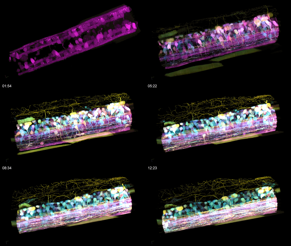
The in vivo development of neural circuitry in the spinal cord of a zebrafish embryo was imaged using the technique. The imaged volume spans 60 x 224 x 180 μm and was acquired over 12 hours. Newly differentiated neurons are highlighted using Autobow fluorophore labeling while older neurons are labeled with mCherry (magenta). The development of rostrocaudally projecting axons can be seen over time across the bottom.
Source: https://goo.gl/VAocpQ ( Science )
Learn More: https://goo.gl/XHYSst (HHMI)
#ScienceGIF #Science #GIF #Biology #Microscopy #Microscope #Zebrafish #Embryo #Spinal #Cord #SpinalCord #Fluorescence #Fluorescent #Lattice #LightSheet #Light #Sheet #SLM #Mirror #2P #Super #Resolution #Modulation #Betzig View Original Post on Google+

