A new microscope developed at Howard Hughes Medical Institute’s Janelia Research Campus is pushing the limits of optical imaging of living organisms. The system combines adaptive optics , a technique commonly used in astronomy to eliminate distortions, with lattice light-sheet microscopy to produce high-speed, super-resolution imagery of cellular dynamics.
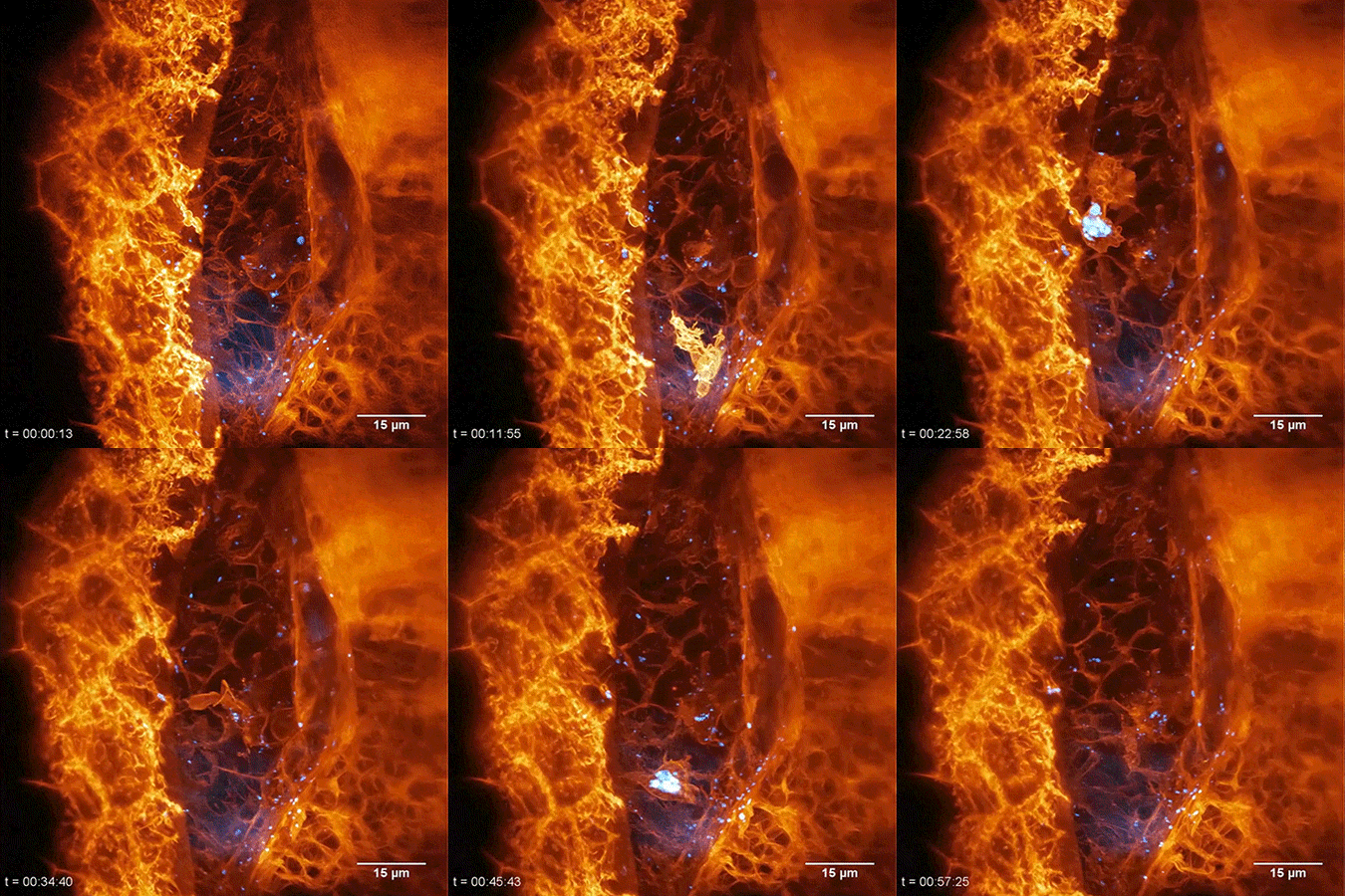
Here, a neutrophil (white blood cell) can be seen internalizing small granules of sugar via phagocytosis within a developing zebrafish embryo. The larva has been genetically modified to express the fluorescent protein Citrine in the plasma membrane of all its cells. Dextran particles labeled with the fluorophore Texas Red were injected into the blood stream and then imaged as they prompted an immune response.
This extraordinary view of cellular behavior within a living organism is one of many remarkable videos reported in Science last week alongside the new microscope.
Source: https://goo.gl/VAocpQ ( Science )
Learn More: https://goo.gl/XHYSst (HHMI)
#ScienceGIF #Science #GIF #Biology #Microscopy #Microscope #Zebrafish #Embryo #Neutrophil #Immune #Cell #Phagocytosis #Sugar #Dextran #Fluorescence #Fluorescent #Lattice #LightSheet #Light #Sheet #SLM #Mirror #2P #Super #Resolution #Modulation #Betzig View Original Post on Google+

