Major advances in optical microscopy over the past decade have allowed for significant breakthroughs in the real-time 3D visualization of biological processes. First demonstrated in 1993, light sheet fluorescence microscopy (LSFM) uses perpendicular illumination of a sample with a light sheet , which is a beam of light focused only in one direction. This allows for the simultaneous illumination of an entire plane in the sample and a significant decrease in acquisition time compared to traditional raster-based techniques.
Lattice light sheet microscopy is an enhanced version of LSFM created in 2014 by Nobel Laureate Eric Betzig that offers even higher spatial and temporal resolutions with reduced photobleaching. The illuminating light sheets are spatially modulated to create 2D optical lattices, which are then scanned across the sample. This allows the technique to break the diffraction limit and achieve super-resolution imaging of living biological samples.
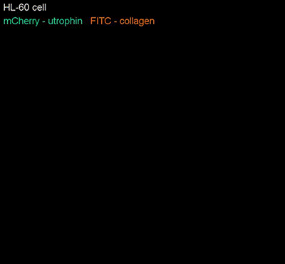
Here, a neutrophil (in green) expressing the fluorescent protein mCherry can be seen navigating through a collagen matrix (in orange) labeled with the fluorophore FITC.
Source: https://youtu.be/UmxKxpKua2M (HHMI)
Journal Article: https://goo.gl/JRuiu7 (Science)
#ScienceGIF #Science #GIF #Cell #Collagen #Matrix #Microscopy #Microscope #Light #Sheet #Lattice #Betzig #Janelia #HHMI #Biology #3D #Neutrophil #HL60 #mCherry #FITC #Optics #Imaging #Super #Resolution #Temporal #Spatial #Modulation #Technology