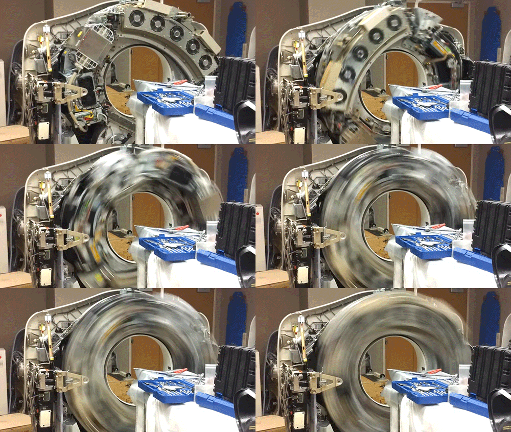
X-Ray Computerized Tomography (CT) produces cross-sectional images of patients by taking X-ray images from different angles and merging them together using a computer. In order to acquire the imagery from different angles, the scanner is rapidly rotated around the patient. Here, the cowling of a CT scanner has been removed to reveal the collection of hardware that is rotated around the central bore hole where the patient is placed.
The CT scanner is comprised of two primary components: the X-ray source and the X-ray detector. The two are located directly across from one another so that X-rays passing through the patient are captured by the detector. Rather than emit a linear beam of X-rays, the source typically emits a fan-shaped beam so that a larger volume of the patient can be imaged. This requires the use of a linear detector array (component with the 5 cooling fans) that captures the expanding beam of X-rays.
Source: https://youtu.be/cjtHNxf01tQ
#ScienceGIF #Science #GIF #CT #CAT #ComputerizedTomography #Medical #MedicalImaging #XRay #Computer #Hardware #Imaging #Technology View Original Post on Google+

